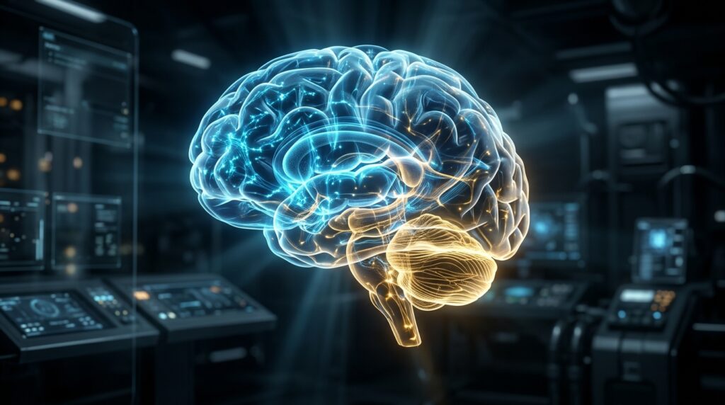
For more than a century, modern medicine and neurobiology rested upon an unquestioned fundamental dogma: that the human brain develops as a single, contiguous biological organ. From early 19th-century histology to contemporary neuroimaging, textbooks have taught generations of physicians and neuroscientists that our entire central nervous system sprouts seamlessly from a solitary, unified pool of embryonic neural progenitor cells. Today, that foundational assumption has been decisively overturned.
In a landmark investigation published in Nature Neuroscience on September 18, 2026, a research team led by developmental biologists at Stanford Medicine has demonstrated that the human brain does not arise from one developmental lineage. Instead, the brain is a composite structure assembled from two distinct, independently evolved organs that emerge from completely separate embryonic progenitor populations before fusing together into a singular anatomical unit.
Led by senior author Dr. Kyle Loh, associate professor of developmental biology at Stanford Medicine, and co-led by graduate researchers Carolyn “C” Dundes and Rayyan Jokhai, the discovery solves a decades-old enigma that has crippled neurological disease research: why scientists have consistently failed to cultivate mature, functional human brainstem neurons in the laboratory. By unraveling this deep developmental duality, the Stanford team has not only rewritten vertebrate evolutionary history, but also unlocked the first reliable platform to model and fight lethal neurodegenerative disorders like Amyotrophic Lateral Sclerosis (ALS) and Spinal Muscular Atrophy (SMA).
The Centenary Dogma: The Myth of the Monolithic Neural Tube
To grasp the seismic nature of this discovery, one must look at how embryology has viewed the nervous system since the foundational work of Swiss anatomist Wilhelm His in the late 1800s. The traditional model posited that following gastrulation, a specialized strip of ectoderm folds inward to form the neural tube. Under this classical framework, every neural structure—from the frontal cerebral cortex responsible for abstract mathematics to the brainstem circuits regulating cardiac rhythm—was presumed to branch from a uniform stem cell pool, patterned along a simple anterior-to-posterior gradient of chemical morphogens.
Yet behind this elegant textbook model lurked an infuriating laboratory paradox. Over the past two decades, the rise of human induced pluripotent stem cell (iPSC) technology enabled researchers to rapidly generate cortical neurons, hippocampal circuits, and retinal organoids with astonishing fidelity. But whenever neurobiologists attempted to produce the neurons of the hindbrain—the vital region comprising the pons, cerebellum, and medulla oblongata—their experiments collapsed into dysregulated, immature, or mispatterned cellular states.
“For years, stem cell biology treated the brainstem like a stubborn child of the forebrain—assuming that if we simply adjusted the concentrations of signaling molecules like retinoic acid and Wnt, we could force embryonic cells into hindbrain fates. What our data prove is that we were trying to speak French to a cell that only understands Mandarin. The hindbrain is not a continuation of the forebrain; it is an entirely separate biological lineage with its own distinct developmental grammar.”
— Dr. Kyle Loh, Associate Professor of Developmental Biology, Stanford Medicine
Otx2 vs. Gbx2: The Molecular Divide of Two Ancient Lineages
Deploying high-resolution single-cell multiomics, epigenomic profiling, and lineage tracing, the Stanford team tracked the earliest topological bifurcation of human pluripotent stem cells during embryogenesis. What they mapped was not a gradient, but an immediate and mutually exclusive genetic crossroads governed by two master regulatory transcription factors:
- The Rostral Lineage (Otx2-Positive): This population exclusively gives rise to the forebrain (telencephalon, cerebral cortex, basal ganglia) and the midbrain (mesencephalon). These structures govern higher cognition, consciousness, executive functioning, emotional processing, and sensory computation.
- The Caudal Lineage (Gbx2-Positive): This distinct population exclusively generates the hindbrain (rhombencephalon, pons, medulla oblongata) and seeds the motor circuits of the upper spinal cord. These structures regulate autonomous vitality—cardiorespiratory pace, involuntary swallowing, motor coordination, and basic autonomic reflexes.
Critically, the Stanford researchers revealed that the divergence between the Otx2 and Gbx2 lineages occurs much earlier than previously imagined—at the very inception of neural specification. The two cell populations exhibit radically different chromatin architectures. In Otx2 progenitors, genomic loci governing brainstem identities are tightly packaged in repressive heterochromatin, rendering them structurally inaccessible. Conversely, Gbx2 progenitors possess a pre-configured epigenetic landscape that permanently prevents them from adopting cortical fates.
Once an embryonic cell commits to either the Otx2 or Gbx2 lineage, its fate is irreversibly sealed. The brain, rather than developing as an unbroken continuum, is built like an architectural bridge where two completely disparate structural foundations are driven into the ground separately before meeting in the middle.
An Evolutionary Merger: Two Ancient Nervous Systems Glued Together
Why would nature construct the crown jewel of human biology out of two independent pieces? The answer lies buried in more than 500 million years of chordate evolution.
Comparative genomic analyses suggest that primitive bilaterian ancestors originally possessed independent neural networks specialized for distinct environmental challenges:
- The Ancient Somatomotor Engine: A primordial caudal nerve center focused on locomotion, rhythmogenesis, respiratory/circulatory control, and visceral reflexes (the evolutionary precursor of the hindbrain).
- The Sensory-Navigational Crown: A rostral sensory ganglion cluster optimized for chemical sensing, light detection, spatial orientation, and adaptive decision-making (the evolutionary precursor of the forebrain).
Over evolutionary epochs, as vertebrate organisms evolved larger body plans, predatory lifestyles, and complex motor repertoires, natural selection literally pushed these two modular nervous systems together. The resulting fusion created a unified central nervous system housed inside the cranium, while preserving the ancient, irreconcilable developmental mechanisms that governed each module’s birth.
Culturing the Unculturable: True Human Hindbrain Motor Neurons
The practical implications of this conceptual paradigm shift are breathtaking. Armed with the realization that the hindbrain follows an autonomous Gbx2-mediated transcriptional path, Dundes, Jokhai, and Loh systematically eliminated forebrain-inducing morphogens from their culture protocols.
Instead, they developed a novel, chemically defined differentiation cocktail that selectively activates the Gbx2 regulatory circuit while suppressing ectopic signaling. The results were immediate and unprecedented:
- Purity Exceeding 92%: The Stanford protocol achieved greater than 90% differentiation efficiency into genuine, bona fide human hindbrain motor neurons, bypassing the messy cellular heterogeneity that plagued prior attempts.
- Electrophysiological Maturity: Using multielectrode arrays (MEAs) and patch-clamp electrophysiology, the cultured motor neurons exhibited spontaneous synchronous burst firing, robust action potential kinetics, and healthy neuromuscular junction formation in co-culture with human skeletal muscle fibers.
- Cranial Nerve Specification: The team succeeded in generating specific sub-classes of motor neurons, including those that form the hypoglossal, vagus, and facial motor nuclei—circuits essential for human speech, facial expression, and breathing.
A Clinical Turning Point: Transforming ALS and SMA Drug Discovery
The inability to culture authentic human hindbrain neurons has been one of the central roadblocks in clinical neurology. Fatal motor neuron disorders do not attack the brain uniformly; they exhibit brutal anatomical selectivity:
In Amyotrophic Lateral Sclerosis (ALS), the disease frequently strikes the bulbar motor neurons situated in the brainstem, leading to rapid loss of speech, dysphagia (inability to swallow), and fatal diaphragmatic respiratory arrest. For decades, drug discovery for bulbar ALS relied on animal models or generic spinal motor neurons derived from flawed forebrain-biased protocols. Consequently, over 99% of prospective ALS neuroprotective compounds failed when brought into human clinical trials.
With Stanford’s breakthrough, biotechnology and pharmaceutical laboratories now possess the exact human cellular canvas required to test interventions:
- High-Throughput Bulbar ALS Screening: Patient-derived iPSCs carrying mutations in SOD1, C9orf72, or TDP-43 can now be differentiated into authentic bulbar motor neurons to screen small molecules and antisense oligonucleotides (ASOs) directly against brainstem-specific neurodegeneration.
- Targeted Cell-Replacement Therapies: The production of pristine, clinical-grade hindbrain progenitor cells opens realistic horizons for regenerative cell therapies aimed at halting respiratory failure in end-stage ALS and Spinal Muscular Atrophy patients.
- Deconvoluting Brainstem Malformations: Pediatric conditions involving congenital hypoventilation (Ondine’s curse) and Chiari malformations can finally be modeled in patient organoid platforms to pinpoint molecular etiology during early trimester development.
Summary: Rewriting the Neurology Textbooks
The Stanford discovery represents more than a technical triumph in stem cell protocol design—it is a philosophical reckoning for medicine. For over a century, neurobiology saw unity where nature had enacted a brilliant modular partnership. By recognizing that our skull houses two ancient, evolutionary sisters working in exquisite synchrony, science has unlocked the keys to protecting them when disease threatens to tear their connection apart.